Introduction
Magnetic resonance imaging (MRI) is renowned for its high-contrast resolution, making it an essential tool in medical diagnostics. The high-contrast resolution of MRI is due to its ability to differentiate between different tissue types based on their water content and relaxation properties [1,2].
However, MRI suffers from relatively long scanning times to acquire high-resolution images. These long scanning times can be attributed to several factors, including the need for multiple sequences, high-resolution requirements, and patient cooperation [3,4].
Long waiting lists for MRI scans are a significant issue in both the USA and Europe, leading to delays in diagnosis and treatment. This problem is multifaceted, involving factors such as the high demand for MRI services, limited availability of MRI machines, staffing shortages, and financial constraints.
Reducing MRI scanning times has become a critical area of focus in medical imaging, aiming to enhance patient comfort, reduce motion artifacts, and increase the throughput of MRI machines. Advances in software and algorithm development have significantly contributed to reducing MRI scanning times. Partial Fourier imaging reconstructs the full image from partial k-space data, reducing the scan time by acquiring only a fraction of the data required for full Fourier reconstruction [8]. Parallel imaging (PI) techniques such as SENSE (sensitivity encoding) and GRAPPA (generalised autocalibrating partially parallel acquisitions) use phased-array coils to simultaneously capture different parts of the image, thus reducing acquisition time [5,6]. Introduced more recently, compressed sensing (CS) technique reconstructs images from under-sampled data by exploiting the sparsity of the image in a certain transform domain (e.g. wavelet domain). It significantly reduces the amount of data needed, thereby shortening scan times [7].
Most recently, artificial intelligence (AI)-based algorithms, particularly deep learning models, are being used to reconstruct high-quality images from significantly fewer data points. These algorithms can predict missing information, thereby reducing the need for extensive data acquisition. In this settings, different vendors including GE Healthcare, Siemens Healthineers, and Philips have developed AI-based MRI acceleration techniques [9].
The development of these advanced software techniques has significantly enhanced the efficiency of MRI scanning, making it possible to obtain high-quality images in a fraction of the time previously required. These technologies continue to evolve, driven by ongoing research and the increasing integration of AI and machine learning into medical imaging. By reducing scan times, these innovations improve patient experience, increase scanner utilisation, and potentially shorten waiting lists.
Reducing MRI scanning offers several advantages, both for patients and for healthcare providers. One of the most intuitive advantages is improved patient comfort. Faster scans mean patients spend less time in the MRI machine, which can reduce anxiety and discomfort, especially for those with claustrophobia. Reduced scan times also lower the likelihood of patient movement, which can degrade image quality and necessitate rescans, making MRI more feasible for those who have difficulties remaining still for long periods, such as children, elders, and people with specific medical conditions. Parallelly, the need for sedated exams could be reduced in clinical practice.
Another key point is the increased accessibility, where quicker appointments can lead to more available slots, reducing waiting times for patients needing urgent diagnostics. Also, shorter scan times allow more patients to be scanned, improving MRI throughput with subsequent better resource management, potentially reducing the need for additional MRI machines and associated costs. These improvements lead to cost-effectiveness: the reduction of operational costs, as shorter scans reduce power consumption, potentially lowering operational and maintenance costs.
On the other hand, deep learning (DL) and AI techniques can be used to increase image quality by reducing the risk of motion artifacts, leading to sharper images and more accurate diagnosis. Also, deep learning reconstruction (DLR) techniques can enhance image quality by effectively reconstructing images from reduced data, potentially providing better diagnostic information even with shorter scans.
From a scientific point of view, faster scanning protocols can accelerate the pace of clinical trials by enabling quicker imaging processes, thereby speeding up research and development timelines. For example, larger datasets can be achieved in shorter times, which can be invaluable for research and the further training of AI algorithms.
Among real-world applications, several fields can take advantage of shortened protocols, which can impact significantly routine exams. In oncologic imaging, faster MRI scans can facilitate more regular monitoring of cancer patients, allowing for timely adjustments to treatment plans. In patients suffering from neurological disorders, rapid brain imaging can aid in the quick assessment of conditions like stroke, multiple sclerosis, and epilepsy, improving patient outcomes. In chronic inflammatory disease, faster imaging may help in reducing the interval between imaging to better check therapy outcomes. All these patients are usually imaged with MRI because of its higher contrast resolution in soft tissues.
Additionally, reducing the scanning time could help MRI to play a role in emergency medicine and acute conditions such as trauma or acute ischaemic stroke. Finally, with short MRI studies available, more routine screenings and preventative health studies could be achieved.
Implementing DLR to reduce MRI scanning times presents multiple advantages, enhancing the patient experience, improving operational efficiency, and pushing the boundaries of medical research and diagnostics. These innovations promise to make MRI more accessible, time-effective, and diagnostically powerful. Probably, within a few years, all MRI scanners will use DLR, which can drastically modify their protocols. The dilemma for radiologists in choosing the level of acceleration for MRI images using DLR lies in balancing image quality, diagnostic accuracy, and scanning efficiency.
In this paper, all the pros and cons of using DLR in clinical practice will be discussed. Additionally, we will share our direct experience of approximately 20 months using dedicated software for reducing acquisition times, installed on the MRI and used at various acceleration factors in clinical routine, to acquire scans of any type, from head to toe.
Scanner and software employed
Image acquisition
All patients were examined with a 3.0 Tesla MR scanner (uMR Omega, United Imaging Healthcare, Shanghai, China). Institutional review board permission was waived for this narrative review study. All data were collected as part of routine care, including only adult patients.
Image reconstruction
All images were reconstructed using conventional acceleration techniques as well as the FDA-approved AI-assisted compressed sensing (ACS). ACS is a DLR that was trained with over 2 million fully sampled slices previously acquired with phantom (2%) and volunteers (98%) [9] (Figure 1).
Figure 1
ACS reconstruction pipeline. ACS integrates specialised mathematical constraints with AI to achieve reliable results. The ACS module effectively remedies reconstruction errors in parallel imaging and half Fourier method, while the reliability issues associated with deep learning’s “black box” effect are solved by the mathematical iterative model, which includes compressed sensing, phase constraints, and data fidelity
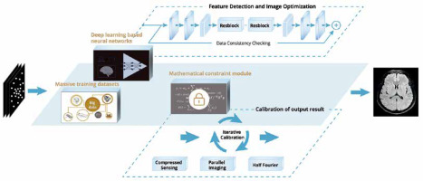
Conventional acceleration techniques combine different acceleration techniques in series like half Fourier (HF), PI, or CS. However, the sequential acceleration structure may lead to error amplification at each level, which threatens data accuracy. ACS provides a new acceleration solution for MRI by innovatively introducing an AI module based on deep neural networks. The output of the AI module is one of the constraint terms for the subsequent iterative reconstruction processes, combined with HF, PI, and CS, to achieve multi-directional image data verification and realise high image quality at ultra-high acceleration.
In ACS, the output image from the reconstruction neural network is subjected to iterative optimisation based on k-space raw data. To ensure trustworthy DL results, a specialised mathematical constraint module is implemented after the net chain, demonstrating the importance of leveraging data information for optimisation in iterative reconstruction, compared to techniques that rely solely on AI performance. Consequently, the AI-enabled, ultra-fast, high-fidelity, and comprehensive whole-body MR application made possible by this technology has given MRI unprecedented degrees of freedom and potential.
ACS is delivered with a graphics processing unit (GPU) based AI reconstruction engine that supports ultra-high-speed “real-time” reconstruction. GPU provides a powerful and reliable hardware support for the training, deployment, and testing of artificial neural network models, with reconstruction speed increasing by more than 43%.
Using deep learning and artificial intelligence in clinical practice
Increase image quality
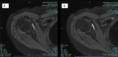
DL and artificial intelligence can enhance imaging quality working on multiple levels [10-13]. Moreover, DLR can produce high-quality images faster while effectively reducing noise in MRI images. By training on large datasets of noisy and clean image pairs, DLR distinguishes and removes noise while preserving important anatomical details. This leads to clearer and more diagnostically useful images (Figure 2).
Figure 2
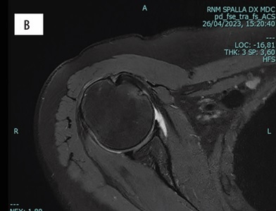
A 46-year-old man undergoing shoulder MRI. On standard axial PD fat saturated image, some noise and blurring artifacts cause a decrease in image quality. The sequence, repeated with a low acceleration factor, significantly improved the image quality, reducing noise and increasing the spatial and contrast resolution
Additionally, super-resolution techniques involve training DLR to reconstruct high-resolution images from low-resolution acquisition. This enhances the detail and clarity of the images, making it easier to identify small structures and subtle abnormalities.
Also, DLR can improve contrast in MRI images by reducing noise level, making it easier to differentiate between tissues with similar signal intensities. This is particularly useful in identifying lesions, tumours, and other pathological features that may not be easily distinguishable with standard imaging techniques.
Artifact reduction
MRI images are prone to various artifacts, such as motion artifacts, Gibbs ringing, and aliasing. DLR can learn to identify and reduce these artifacts, resulting in even cleaner images. For example, models can be trained to recognise patterns associated with motion and adjust the images accordingly [14-17].
Johnson et al. [16] presented a conditional generative adversarial network (GAN) approach to correct for rigid-body motion artifacts in 3D MRI, showing significant improvements in image quality. In another recent paper, Tamada et al. [18] explored a DL framework designed to reduce motion artifacts in MRI, comparing its performance to traditional techniques and demonstrating enhanced image clarity and diagnostic value.
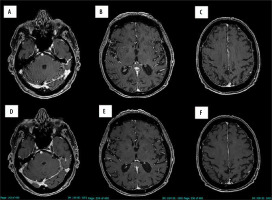
Simply but very effectively, motion artifacts can be reduced or even eliminated in difficult patients by significantly reducing acquisition times. For example, by halving the scan times for uncooperative patients, it is possible to remove annoying motion artifacts in the brain, achieving excellent image quality (Figure 3).
Figure 3
A 58-year-old man suffering from involuntary spasms, undergoing brain MRI. Axial post-contrast-enhanced T1 images show heavy motion artifacts on different levels (A, B, C). The sequence, repeated with a medium acceleration factor, allowed significant improvement of the image quality by eliminating the motion artifacts (D, E, F)
By leveraging DLR to accelerate MRI scans, enhance image quality, and reduce motion artifacts, the overall MRI experience can be made more tolerable for patients, decreasing the need for sedation. This is particularly beneficial for vulnerable populations such as children, elderly patients, and patients with conditions that make staying still difficult.
The key factors are the acceleration of scan times, improving image quality and reducing motion artifacts. Shorter and more efficient scans can help reduce the anxiety and discomfort associated with MRI procedures, particularly for paediatric patients, thus reducing the reliance on sedation.
Automated patient positioning
Automated patient positioning in MRI is a technological advancement that aims to improve the accuracy and efficiency of MRI exams [19,20]. This technology utilises AI and machine learning algorithms to position patients correctly within the MRI scanner, thereby reducing setup times and minimising operator-dependent variability. Automated positioning systems can standardise the patient setup process, ensuring consistent and optimal positioning for every scan. This reduces the likelihood of rescans due to suboptimal positioning, which can save time and improve patient throughput.
These systems typically use DL algorithms trained on large datasets of MRI scans to recognise anatomical landmarks and position patients accurately. The algorithms can adapt to various body types and scanning requirements, further enhancing the reliability of the positioning.
By automating the positioning process, technologists can reduce the time spent on manual adjustments, allowing for a quicker transition between patients and more efficient use of the MRI scanner.
Automated positioning can be particularly beneficial in settings where high patient throughput is required, such as in large hospitals or imaging centres. It can also be advantageous for paediatric or geriatric patients, who may have difficulty remaining still for extended periods.
Reducing the scanning time
AI and DL play a pivotal role in reducing scanning times in MRI by optimising various aspects of the imaging process. DLR uses a combination of techniques such as CS and PI. Traditional MRI relies on fully sampling k-space data, but DLR can reconstruct high-quality images from under-sampled data. Recent AI driven software can work both during the acquisition of data, and during the reconstruction of images. GANs can generate high-resolution images from lower-resolution inputs, effectively reducing the need for long scanning times [21]. Also, AI can lead to the use of smart protocols: in particular, AI-driven protocols can optimise scanning protocols by selecting the best sequences and parameters tailored to each patient, thus shortening the overall scan time [22].
Reducing redundant data acquisition by using an adaptive sampling approach is another way AI can work. AI can adaptively decide which parts of k-space need to be sampled more densely and which can be under-sampled, reducing the overall acquisition time without sacrificing image quality [23].
Enhanced image quality, by means of noise reduction to enhance the signal-to-noise ratio of MR images, allows for shorter acquisition times because fewer data are needed to achieve the same image quality [24].
Recently, a patient-specific scanning strategy (tailored imaging) is based on models that predict patient-specific anatomy and pathology, allowing for more efficient scanning strategies tailored to the individual’s needs.
AI and DL are revolutionising MRI by enabling faster, more efficient scans through advanced reconstruction techniques, real-time artifact correction, optimised protocols, adaptive sampling, and enhanced image quality. These innovations reduce the time patients spend in scanners, improve patient comfort, and increase the throughput of MRI facilities. Shorter scan times allow for quicker diagnosis, enabling timely treatment initiation which can be crucial for conditions like stroke, trauma, and certain cancers [25].
One of the most important achievements is the increased throughput: MRI facilities can accommodate more patients per day, reducing waiting times and improving access to necessary imaging services.
As a consequence, an optimised workflow could be allowed by shorter scan times that can streamline workflow in radiology departments, allowing for better resource management and reduced bottlenecks [26]. Also, the increased efficiency and patient throughput can lead to lower operational costs and better utilisation of expensive MRI equipment. Among improvements in clinical outcomes, the possibility of enhanced follow-up due to easier access and reduced scan times can facilitate more frequent monitoring of chronic conditions and hence improve the effectiveness of treatments.
The issue of diagnostic accuracy in clinical practice
The integration of DL into MRI sequences has shown promise in maintaining, or even improving, diagnostic accuracy while reducing scanning times [23]. Some clinical validation studies have validated the clinical efficacy of DLR MRI protocols. These studies involve comparing the diagnostic performance of conventional MRI sequences with those accelerated by DLR. Johnson et al. [27] conducted a prospective study on 170 patients and found that the diagnostic accuracy of DLR MRI images was comparable to standard protocols, with substantial reductions in scanning times.
DLR MRI has been applied successfully in various clinical scenarios, including brain imaging, musculoskeletal imaging, and cardiac imaging. These applications have shown that DLR can be generalised across different types of MRI exams. Sriram et al. [28] explored the use of DL in musculoskeletal MRI and demonstrated that DLR protocols could achieve high diagnostic accuracy across different anatomical regions.
The radiologist’s concern: How far can I push AI to reduce acquisition time?
With the advent of new MRI scanners, often equipped with AI and DL to reduce acquisition times, radiologists naturally start to ask some fundamental questions. Given that these systems are usually easy to use and should not significantly alter reconstruction times, it remains to be seen whether one should rely on DLR to reduce acquisition times compared to standard sequences. The fear for the average radiologist, accustomed to their workflow, might be losing fine details in the images that could affect their final judgment. MRI diagnoses are becoming increasingly precise, with sub-millimetric morphological alterations and signal changes sometimes so subtle that they can only be visualised with very long scanning times. It is therefore natural for professionals to worry that the quality of their diagnoses might decline. Furthermore, even radiologists used to working with AI and reduced acquisition times might have a significant concern: How far can I push the acceleration factor? How reliable is AI as data under-sampling increases?
It is evident that the literature does not yet provide sufficient answers to these questions. While there are indeed some well-structured prospective studies demonstrating that DLR images do not significantly reduce diagnostic accuracy, these studies are very limited in number and often focused on specific sequences and topics. Therefore, it is not possible to generalise their findings. Additionally, many studies result from using dedicated AI and DL kits rather than proprietary software that radiologists would use in their clinical practice. The hope is that dedicated, possibly multicentre, studies will emerge, prospectively and progressively testing the potential diagnostic accuracy of compressed sequences.
Personal experience
The following considerations arise from over 20 months of field experience using a 3T MRI scanner equipped with clinical product DLR capable of enhancing image quality while maintaining scan times or reducing acquisition times and keeping acquisition parameters unchanged. Because the image quality of standard sequences for any examined region, from head to toe, was considered very high (and certainly superior to the other 1.5 T scanners in our institute), we decided to optimise study protocols to reduce acquisition times and generally increase patient access to the scanner. To this end, we began testing the potential use of DLR scans with reduced times in a fairly random manner, repeating one or more key sequences of the study while keeping the acquisition parameters but increasing the acceleration level. This simple approach was favoured by the ready availability of the software on the scanner and the extreme ease of use (only the acceleration factor needs to be set, and there are typically no delays in reconstruction). After this initial approach, the first results were very promising.
We are not just talking about maintaining good image quality without encountering artifact-laden images, but also about being able to identify the smallest pathological alterations in MRI images, whether they are small anatomical or morphological changes or subtle signal alterations. It seemed that even with significantly reduced acquisition times, we could always see the same findings, allowing for a correct diagnosis every time. However, this brings about a real dilemma for radiologists in choosing the acceleration factor for MRI images reconstructed using DLR. Higher acceleration factors can significantly reduce scan times, making the process quicker and more comfortable for patients. However, excessive acceleration factor may lead to loss of critical details, potentially compromising diagnostic accuracy. Lower acceleration factor maintains higher image quality but result in longer scan times, which can be less efficient. Also, in critical diagnoses such as small lesions, microbleeds, or subtle tissue changes, high-resolution images are crucial.
The diagnostic problem (from the perspective of a radiologist used to state-of-the-art work using non-DLR sequences) is that “artificial” images created by compensating for data under-sampling using DLR might mask or even produce artifacts that do not actually exist. The problem becomes more complex in oncologic patients. In these cases, the presence of a single millimetric metastasis can radically alter staging and thus the subsequent diagnostic and therapeutic pathway. Equally concerning is the possibility of a false positive, especially in oncology, where a patient with a false positive diagnosis of a small metastasis might undergo a drastic and unnecessary change in their management plan.
To address these questions, our institute is currently conducting several studies focused on the use of DLR sequences to reduce acquisition times. These studies share the same methodological framework: clinical studies that fall within the research topic are selected, and the standard protocol is performed without DLR, which will serve both for clinical diagnosis and to create our reference standard for the final diagnosis. Key sequences for the study are then selected and repeated using increasing acceleration factors for DLR (Figures 4 and 5). The obtained datasets are then re-evaluated by various readers, and the results are compared with those of the standard exam to calculate diagnostic accuracy.
Figure 4
A 64-year-old woman suffering from breast cancer, undergoing brain MRI. Axial post-contrast-enhanced T1 image (A) identifying a single, tiny, enhancing lesion consistent with the diagnosis of a small metastasis (yellow circle), showing only mild perifocal oedema on corresponding axial reconstructed FLAIR image (yellow arrow on D). The sequences, repeated with a 2-fold acceleration factor (B, E) and with a 3-fold acceleration factor (C, F), confirm the presence of this tiny lesion (circle and arrow) while maintaining good image quality
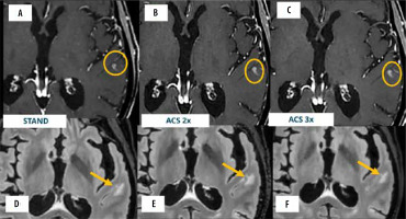
Figure 5
Testing high acceleration factor on PD fat sat imaging of the knee. A 42-year-old man suffering from lateral knee pain undergoing knee MRI. Standard coronal PD FAT sat image (A) identifying a large tear of lateral menisci (arrow). The same meniscal tear (arrow) is confirmed on the 2-fold (B) and 4-fold accelerated images (C), while maintaining good image quality. On the corresponding 6-fold accelerated image (D), the meniscal tear is still beautifully depicted (arrow) despite a relative drop of image quality

Currently, preliminary results from ongoing studies demonstrate diagnostic accuracy values of around 99% for medium-high compression levels (2-3×) and nearly 100% for low compression levels. The results of these studies will be used as an internal reference for the optimisation of daily protocols and may provide a methodological reference for other authors and radiologist colleagues in their daily practice.
At present, based on preliminary results, our institute’s approach would involve the following steps.
For routine scans where fine details are less critical, higher acceleration factor levels might be acceptable, facilitating faster scans and higher patient throughput. This is particularly true for anatomical imaging that might tolerate higher acceleration factors without significant loss of diagnostic information.
For complex studies, we prefer to use very low acceleration factors, which can still result in significant time savings on sometimes very long sequences (e.g. a nonaccelerated sequence of 10 minutes can be reduced by 30% to 7 minutes). This is the case of functional studies like functional MRI (fMRI) or diffusion tensor imaging (DTI).
Also, in daily practice in cases where patient cooperation is limited (e.g. paediatric patients or those with movement disorders), faster scans with higher acceleration factors might be preferred, to reduce motion artifacts. Similarly, in emergency situations, the need for rapid diagnosis might outweigh the need for the highest image quality, prompting the use of higher acceleration factors (e.g. stroke imaging).
The use of DL and AI in clinical practice will bring new challenges for radiologists. Radiologists must be familiar with how DL algorithms affect image quality at various acceleration factors. This requires additional training and experience with the technology.
There is no universal standard for the optimal acceleration factor. Different manufacturers and institutions may have varying recommendations and thresholds, making it challenging for radiologists to make consistent choices.
Radiologists must consider a multitude of factors simultaneously, including the specific clinical question, the anatomical area being imaged, and the patient’s condition, which can complicate the decision-making process.
Finally, the performance of DLR algorithms can vary between different MRI machines and software versions, introducing another layer of complexity in choosing the appropriate acceleration factor.
Potential future solutions could be the basis of clinical guidelines. Development of clinical guidelines and protocols that provide recommendations for acceleration factors based on specific clinical scenarios and anatomical regions can help standardise decisions and reduce variability.
Conclusions
In summary, reducing MRI scan times through the implementation of AI and DL technologies can lead to a range of benefits, including improved patient comfort and experience, enhanced diagnostic efficiency, operational benefits for healthcare providers, better clinical outcomes, reduced motion artifacts, and expanded access to imaging services. These advantages collectively contribute to a more effective and efficient healthcare system.


