Introduction
Congenital heart diseases (CHD) form an important spectrum of paediatric diseases causing significant morbi-dity and mortality. Due to inter-related embryology of the heart and development of the coronary arteries, there is great variability in coronary artery patterns in patients with CHD [1]. It is critical to evaluate the origin and course of the coronary arteries when planning surgical or interventional therapies in patients with CHD [2]. The presence of an anomalous origin or course of a coronary artery can lead to a modification in surgical approach to a particular disease or anomaly of the heart, and it can pose a danger of inadvertent damage to the coronary arteries during surgery. These variations also influence the outcomes of surgical procedures for various complex congenital heart surgeries [3-5].
Due to rapid technological advancements and increased speed of acquisition with improved spatial and temporal resolution, multidetector computed tomography (MDCT) plays a critical role in the diagnostic evaluation of children with suspected or diagnosed CHD [6-8]. Due to the acquisition of an isotropic dataset, high-quality multiplanar reformation, curved reformats, and three-dimensional (3D) reconstructions including maximum intensity projections (MIP) and volume-rendered techniques (VRT) are now possible. Detailed assessment of coronary anatomy origin, course, and termination, its lumen, and the vessel wall can be done. Despite these advantages, there is a risk of radiation exposure with CT [5]. However, several new proposed sets of factors, including high pitch, optimization of kilovoltage and tube current, prospective electrocardiogram (ECG) gating, and use of iterative reconstruction, can reduce the radiation dose to a mini-mum [9,10].
Due to higher heart rate in young children and uncooperative respiration, coronary artery evaluation with non-ECG-gated studies is difficult [11]. With ECG gating, there is a significant improvement in coronary artery visualization in children and infants. Improved detection rate, accuracy, and image quality have been associated with ECG-gated rather than non-ECG-gated techniques in the evaluation of coronary arteries in children [12]. Furthermore, retrospective ECG-gated studies incur more radiation as compared to prospectively gated studies [13]. MRI studies are still not routinely performed and are considered inferior to CT scans, specifically for coronary artery visualization in children [14].
We conducted this retrospective study to compare the visualization and anatomical variations of coronary artery diseases in children with CHD on non-ECG-gated and ECG-gated CT angiography studies.
Material and methods
Study population
We retrospectively identified consecutive patients ≤ 2 years of age who underwent single-phase multidetector computed tomography (MDCT) thoracic angiography for anatomical and morphological assessment of suspected or confirmed CHD without (January 2020 – November 2020) and with (December 2020 – December 2021) ECG gating.
Forty children underwent a non-ECG-gated study (group A), and 42 children underwent an ECG-gated study (group B) during the study period.
This retrospective study was approved by the Ethics Committee of our institute, and a waiver for informed written consent was obtained. Patient confidentiality was maintained in accordance with Health Insurance Portability and Accountability Act guidelines.
Computed tomography angiography acquisition
All the children underwent single-phase acquisition using the bolus tracking technique from the lung apices to domes of the diaphragm using non-ionic iodinated contrast medium (300 mgI/ml) with a contrast dose of 2 ml/kg using a power injector with the rate based on the size of intravenous cannula. All the studies were obtained with weight-based kilovoltage and low-dose tube current.
Non-electrocardiogram-gated multidetector computed tomography studies
The non-ECG-gated MDCT thoracic angiographic studies were acquired on one of the 2 MDCT scanners available, which included Somatom Definition 128-slice (Siemens) and Philips Brilliance iCT 256-slice (Philips Healthcare) scanners from January 2020 to November 2020, before the installation of the Somatom Force CT (Siemens) at our institute.
Electrocardiogram-gated multidetector computed tomography studies
The ECG-gated thoracic MDCT angiographic studies were acquired on a Somatom Force CT (Siemens) scanner from December 2020 to December 2021. All the studies were prospectively gated with turbo flash spiral data acquisition. Automated selection of mAs (milliampere-seconds) was done with CARE dose 4D dose modulation software, which is inbuilt in the scanner.
The specific thoracic MDCT parameters for the 3 different MDCT scanners used are summarized in Table 1.
Table 1
Acquisition parameters for the 3 different multidetector computed tomography scanners
Multidetector computed tomography image reconstruction
The axial MDCT images were reconstructed at a submillimetre slice thickness for review. Two-dimensional (2D) multiplanar reformat (i.e. sagittal and coronal planes) images were also reconstructed from datasets with submillimetre slice thickness. In addition, three-dimensional (3D) maximum intensity projection (MIP) images (i.e. axial, sagittal, and coronal planes) and volume-rendered CT images were generated.
Image review
Two paediatric radiologists, with 10 and 20 years of experience in interpreting MDCT thoracic angiography studies in children with CHD, independently reviewed all the non-gated and gated MDCT cardiac angiography studies. Any discrepancy in the findings was resolved by consensus.
All MDCT studies were evaluated with standard soft tissue (width, 350 HU; level, 50 HU) window settings. Multiplanar, MIP, and volume-rendered 3D CT images were also used routinely by the radiologists for the evaluation of coronary arteries. The right coronary artery (RCA), left main coronary artery (LMCA), left anterior descending artery (LAD), and left circumflex artery (LCX) were evaluated. All the coronary arteries were evaluated for the visualization of their origin at the aortic sinus and their course. They were classified as visualized when both the origin and course were properly visualized, and as not visualized when either the origin or the course were not properly visualized. In addition, variations in normal coronary anatomy were also noted.
Statistical analysis
Continuous variables that were normally distributed, as assessed with the Kolmogorov-Smirnov goodness-of-fit test, such as age, were expressed as means, standard deviations, and ranges. Interobserver agreement for the 2 independent radiologists regarding MDCT angiography findings in the non-gated and gated groups was determined using the chance-corrected kappa coefficient, where excellent agreement is represented by a k value greater than 0.75. Statistical analysis was performed with the SPSS statistics software package (version 21.0, IBM).
Results
Study population
Multidetector computed tomography scanners
Out of the 40 children in group A, 26 (65%) underwent CT on a Somatom Definition 128-slice scanner and 14 (35%) on a Philips Brilliance iCT 256-slice scanner.
All of the 42 (100%) children in group B underwent CT on a Somatom Force CT.
The mean CTDI in the ECG-gated group was 3.02 ± 1.26 mGy, while it was 9.96 ± 13.38 mGy in the non-ECG-gated group.
Multidetector computed tomography findings
Type of congenital heart diseases
Both cyanotic and acyanotic CHD were encountered in both the study groups. The most common CHD in Group A was aortic arch abnormality, which comprised 32.5% of all the cases, while the most common CHD in group B was tetralogy of Fallot (TOF) morphology, which comprised 33.3% of all cases. Table 2 summarizes the type of congenital heart abnormalities encountered in the 2 groups.
Table 2
Table summarizing the different congenital heart diseases seen in the 2 groups
Coronary arteries evaluation
Right coronary artery
The right coronary artery (RCA) was visualized in 19 (47.5%) patients in group A, while it was visualized in 27 (64.3%) patients in group B. It was not properly visualized either at origin or in its course in 21 (52.5%) patients in group A and in 15 (35.75%) patients in group B.
Left main coronary artery
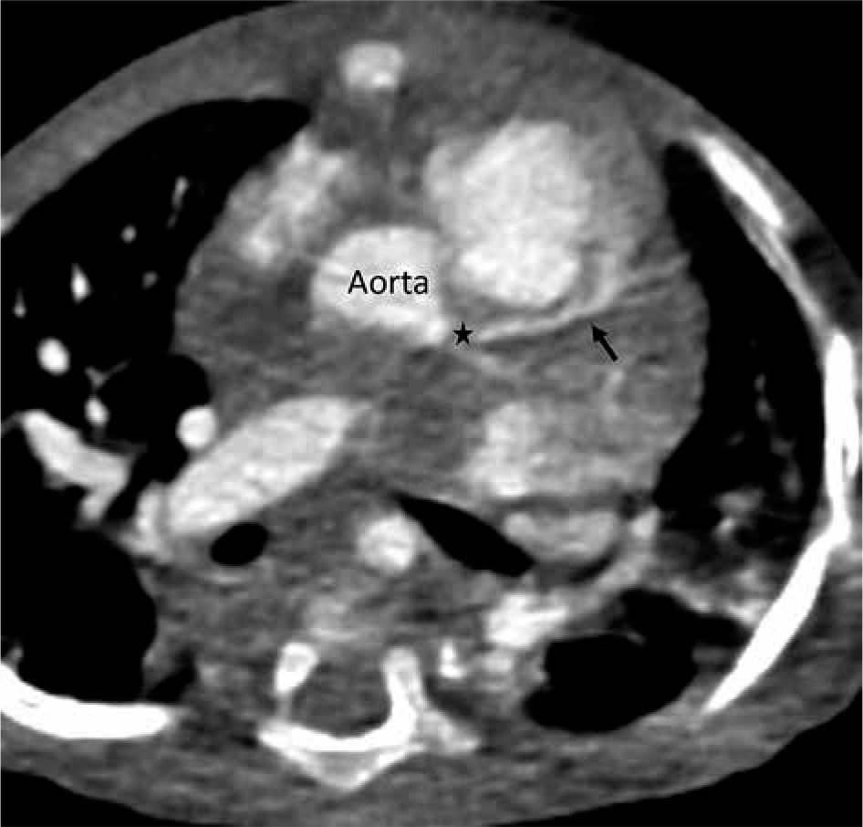
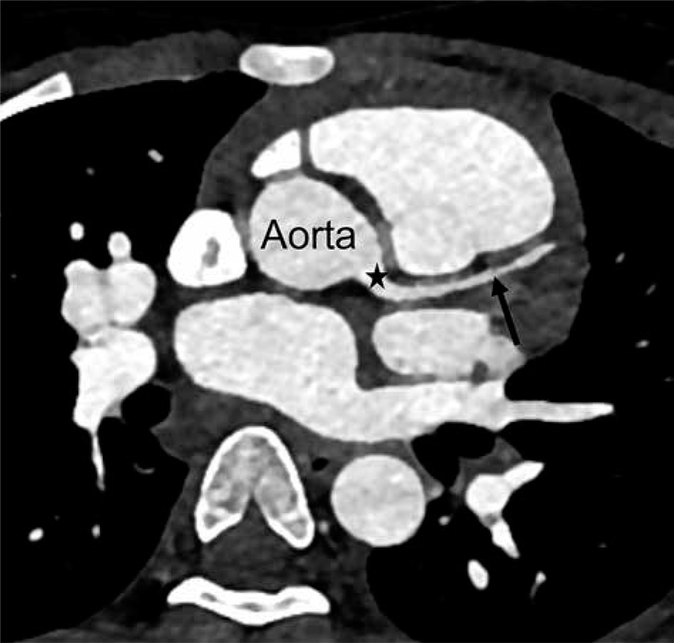
The left main coronary artery (LMCA) was visualized in 25 (62.5%) patients in group A (Figure 1), while it was visualized in 39 (92.8%) patients in group B (Figure 2). It was not properly visualized either at origin or in its course in 15 patients in group A (37.5%) and in 3 patients in group B (7.2%).
Left anterior descending artery
The left anterior descending (LAD) artery was visualized in 22 (55%) patients in group A (Figure 1), while it was visualized in 34 (80.9%) patients in group B (Figure 2). It was not properly visualized either at origin or in its course in 18 (45%) patients in group A and in 8 (19.1%) patients in group B.
Coronary artery variations
Anomalous coronary arteries were identified in only one patient in group A, in the form of a single origin of the RCA and the LMCA.
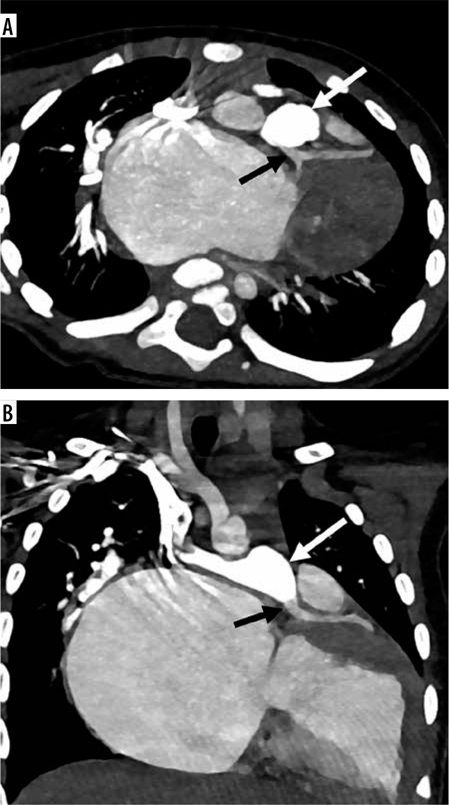
Variations in coronary arteries were identified in 10 (23.8%) patients in group B. Two patients had a common origin of RCA and LMCA. Two children had an early branching of the LAD from the LMCA. In 2 children, the LAD artery was seen arising from the RCA, while in one child the LAD artery was seen arising from the main pulmonary artery (Figure 3). There was an anomalous origin of the LMCA and RCA from the right and the left aortic sinus in 2 children, respectively. In one child, double RCA/splitting of the RCA was seen.
Discussion
In our study of the coronary arteries in children less than 2 years of age, ECG-gated thoracic angiography studies performed significantly better for the detection of all the coronary arteries in comparison to the non-ECG-gated studies. The LMCA, LAD artery, and LCX artery were detected almost 30% more by ECG-gated MDCT versus non-ECG-gated MDCT. The RCA was detected almost 15% more with an ECG-gated study in comparison to a non-ECG-gated study. Also, the coronary artery variations were detected in a significantly higher number in the ECG-gated group (26.2%) than the in non-ECG-gated group (2.5%).
The prevalence of coronary artery anomalies is variable and ranges from 0.85 to 5.6% [10], with a higher incidence (3-36%) in children with CHD [15]. In patients with CHD who are candidates for surgical intervention, delineation of the origin, course, and the termination of coronary arteries is important for surgical planning. The coronary artery course has particularly high variability in patients with transposition of the great arteries (TGA). Inadvertent damage of anomalous arteries can occur if the exact origin and course are not known before the surgery. In patients with TOF, an anomalous artery or a large conus branch may pass over the right ventricular outflow tract and may be severed during ventriculotomy. This may necessitate surgical implantation of a right ventricular-to-pulmonary artery conduit [16]. Coronary artery re-implantation may be needed in arterial switch operations [17].
Imaging of coronary arteries in children is challenging because of complex cardiac motion, smaller size, and tortuous courses of the arteries [18]. With the technical advances in CT imaging, including high pitch factors, prospective ECG gating, iterative reconstruction, and optimized tube voltage and current, there has been an increasing role of CT angiography in young children and neonates with CHD for morphological and detailed anatomical evaluation, which is crucial for appropriate surgical planning [19,20].
Goo et al. [11] detected proximal segments of coronary artery in more than 81.7% of cases; however, their study was performed in older children. They also reported that the LMCA and the proximal portions of the LAD artery were the most visible portions of coronary arteries, which was similar to the results found in our study. This could possibly be due to the straighter course of the LMCA, LAD arteries as compared to RCA and LCX artery, both of which run in the atrioventricular groove. Tsai et al. [12] in their study detected 60% more proximal and distal coronary segments with a ECG-gated MDCT study in comparison to non-ECG-gated study, which was also similar to the findings in our study. Also, better image quality, rate of detection, and accuracy were seen with ECG-gated techniques in comparison to non-ECG-gated techniques. We believe that increased temporal resolution of the scanner with better detector technology and more sophisticated reconstruction techniques might also have contributed to the better image quality and increased visualization of the coronary arteries in our study. Hence, future studies may be performed for the evaluation of coronary arteries on the same scanner with and without the use of ECG gating to validate the findings of our study.
Huang et al. [21] used prospective ECG gating to evaluate CHD in infants and reported that more than 80% of scans were of diagnostic image quality to delineate the coronary arteries. Thus, for neonates with CHD, especially requiring surgery, an ECG-gated CT study should be used. Although non-gated techniques can provide gross morphological information, it is not as good for accurate delineation of coronary arteries. Routine use of ECG-gated angiography studies in children with CHD started after the installation of the Somatom Force CT (Siemens) at our institute. Due to higher radiation dose involved with CT scans, strict ALARA principles should be followed while planning MDCT thoracic angiographic studies [22].
We acknowledge a potential limitation of our study. The patients in the 2 groups in our study were scanned on different scanners, and hence increased temporal resolution of the scanner with better detector technology and more sophisticated reconstruction techniques might also have contributed to better image quality and increased visualization of the coronary arteries. Hence, future studies may be performed for the evaluation of coronary arteries on the same scanner with and without the use of ECG gating, to validate the findings of our study.