Introduction
Recurrent caries under different restorative materials are a major cause of the failure and replacement of restorations [1,2]. Although accurate diagnosis of these caries can lead to successful treatment outcomes, it remains a challenging step for clinicians. To provide better detection, visual and radiographic examinations accompanied by other diagnostic methods play a crucial role in treatment planning [1,3]. For these purposes and considering the high prevalence of recurrent caries, periapical and bitewing radiographs are the most common imaging modalities in clinical dentistry. However, misinterpretation of caries detection using existing conventional and 2D radiographic techniques may occur because of anatomic noise, superimposition of structure, and projection geometry [4].
To overcome the shortcomings of the two-dimensional radiographic method, cone-beam computed tomography (CBCT), with the advantage of three-dimensional assessment of dentomaxillofacial structures and their multiplanar transformations, should be considered as a preferred imaging modality. In fact, the use of the CBCT system provides some benefits, such as easier image acquisition, less space requirements, and various sensor types [5-7]. However, beam-hardening and streak artifacts are known to be limiting factors for the precise detection of caries in CBCT, which are related to the dark areas around materials and metallic structures (e.g. restorations, fillings, implants, and endodontic instruments). Different methods have been proposed for artifact reduction in CBCT scans [8,9]. Thus, fewer metal artifacts in the field of view (FOV) will increase the overall quality of the images while providing higher spatial resolution. Moreover, the CBCT unit and imaging parameters could influence the formation of artifacts [10].
Recent developments offer CBCT scanners with a greater dynamic range and superior technical specifications for detecting recurrent caries. The user’s choice of CBCT scanners varied between high and low spatial resolution settings and different imaging modes, which led to reduced radiation doses and increased image quality [11]. Therefore, it is clinically essential to evaluate the performance of CBCT systems using various resolution and imaging scan modes for the visibility of recurrent caries. In the literature, only one study by Cheng et al. [11] focused on the detection of proximal caries by phosphor plate and CBCT images scanned with different resolutions; the authors suggested that spatial resolution does not significantly affect the diagnostic accuracy of flat panel CBCT images for proximal caries [11].
Until now, there have been few studies comparing different CBCT scan modes with and without the metal artifact reduction (MAR) mode in detecting recurrent caries under various restorative materials [12]. In our previous study, we did not apply the MAR option [13]. To better diagnose recurrent caries, the purpose of the current CBCT-based study was to compare the diagnostic performance of 2 CBCT scan modes (high-resolution [HIRes] and standard) with and without the application of a MAR option under 5 different restorative materials in an in vitro setting. The null hypothesis of no significant differences in diagnostic outcomes was postulated for the CBCT scan modes, types of restoration, and activation of MAR.
Material and methods
Study design
The present in vitro study comprised 150 human premolar and molar teeth that were extracted for periodontal or orthodontic reasons. All selected teeth had a complete crown and a normal anatomical shape and were evaluated using a sharp dental explorer (Fattahteb, Tehran, Iran). Teeth surfaces were cleaned of calculus and debris, sterilised in 0.5% sodium hypochlorite for one week, and stored in distilled water. Approval for the use of extracted human teeth was obtained from the Research Ethics Committee of Babol University of Medical Sciences (ethics code: IR.MUBABOL.HRI.REC.1401.116).
Preparation of specimens
For cavity preparation, a standard deep class II cavity was created in the middle of the mesial surface of teeth using a turbine (Push button, Goldent, China) and 008 fissure bur (Tizkavan, Tehran, Iran). The buccolingual width of the cavity was 3 mm, and the gingival floor was 1 mm below the cementoenamel junction. After preparing the cavity, the teeth were kept in a normal saline solution.
To induce artificial caries, the teeth were randomly divided into 2 groups: an experimental group and a control group, with each group consisting of 75 teeth. The sample size was calculated using the following formula, where P is the average of P1 and P2, and Zα and Zβ are the standard normal Z values corresponding α and β (the probability of type I and type II errors, respectively). This formula is mainly used to compare the sensitivity or specificity of the 2 tests. To determine the sample size, we considered an α value of 5% and a β value of 10%.
In the experimental group of 75 teeth, secondary caries were artificially created with the demineralising solution in the gingival wall, and 75 control teeth were without caries. For the demineralising solution to affect the gingival surface, all tooth surfaces except the gingival floor were covered with 2 layers of acid-resistant nail polish. After 8 weeks, caries with an average thickness of 500 microns were made on the teeth gingival surface, and the nail polish was removed with a bur from the teeth of the experimental group. Caries formation was approved in a pilot study under a polarising light microscope (Leitz sm lux pol binocular polarizing light Microscope).
Each group was divided into 5 subcategories (n = 15), and the teeth were filled with different types of restorative material. The filling steps for both groups are similar. In this study, the following 5 types of restorative materials were utilised for teeth filling: conventional composites (Filtek Z250 [3M, ESPE, United States], Gradia [GC, Tokyo, Japan]), flow composite (A2 Filtek Z350 [3M, ESPE, United States]), glass ionomer (Fuji II LC [GC, Tokyo, Japan]), and amalgam (SDI-GS-80). Finally, the teeth were mounted in 4 rows of plaster blocks to have a proximal contact. The blocks with 4 teeth were placed opposite each other and fixed in occlusion with wax (Figure 1). In total, there were 18 blocks with 8 teeth and one block with 6 teeth.
Figure 1
A) Teeth in plaster blocks (upper, from left to right: composite Filtek Z250 without recurrent caries, composite Filtek Z250 without recurrent caries, composite flow with recurrent caries, composite Gradia with recurrent caries; lower, from left to right: composite amalgam with recurrent caries, composite Filtek Z250 without recurrent caries, composite Filtek Z250 without recurrent caries, composite Filtek Z250 without recurrent caries). B) Imaging the restorative materials using the cone-beam computed tomography (CBCT) scan. C) High-resolution CBCT with metal artifact reduction application. D) Standard CBCT with metal artifact reduction application. E) High-resolution CBCT. F) Standard CBCT
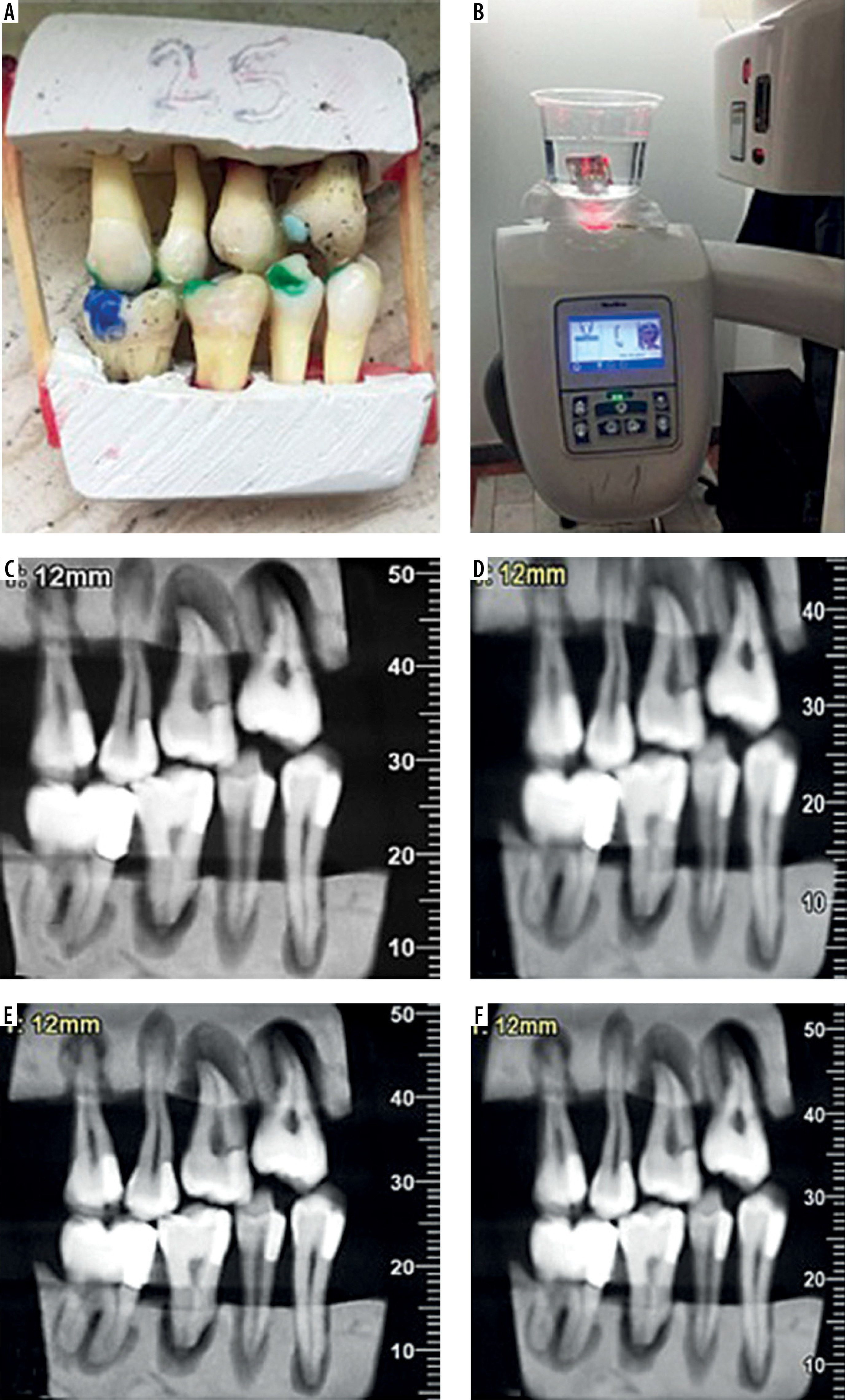
Cone-beam computed tomography imaging
For imaging procedures, all teeth were mounted in plaster blocks, and there was no soft or hard tissue to attenuate radiation. Therefore, to simulate in vivo conditions, the mounted teeth in each mandible were placed in a plastic container containing 1 litre of distilled water. Afterward, the teeth were irradiated and imaged using the CBCT Giano (Newtom, Verona, Italy) system in a private clinic. All images were acquired using 2 different system scan modes as follows: Mode 1: HIRes mode at 90 kVp, 8 × 11 cm FOV, 3 mA, and an exposure time of 9 s using 46.9 µm voxel size; Mode 2: standard-resolution at 90 kVp, 8 × 11 cm FOV, 3 mA, and an exposure time of 3.6 s using 150 µm voxel size, with and without MAR. We selected this specific FOV to ensure accurate imaging of the teeth within the plaster blocks. Our study involved a comprehensive evaluation and comparison of dental caries detection under various restorative materials using CBCT scans, both with and without MAR application. In total, 76 CBCT images (including HIRes mode with/without MAR and standard-resolution mode with/without MAR) were prepared from the 19 blocks.
After image acquisition, the images obtained with NewTom were imported into the NNTTM Viewer software (Newtom, Verona, Italy) and are shown in Figures 2 and 3.
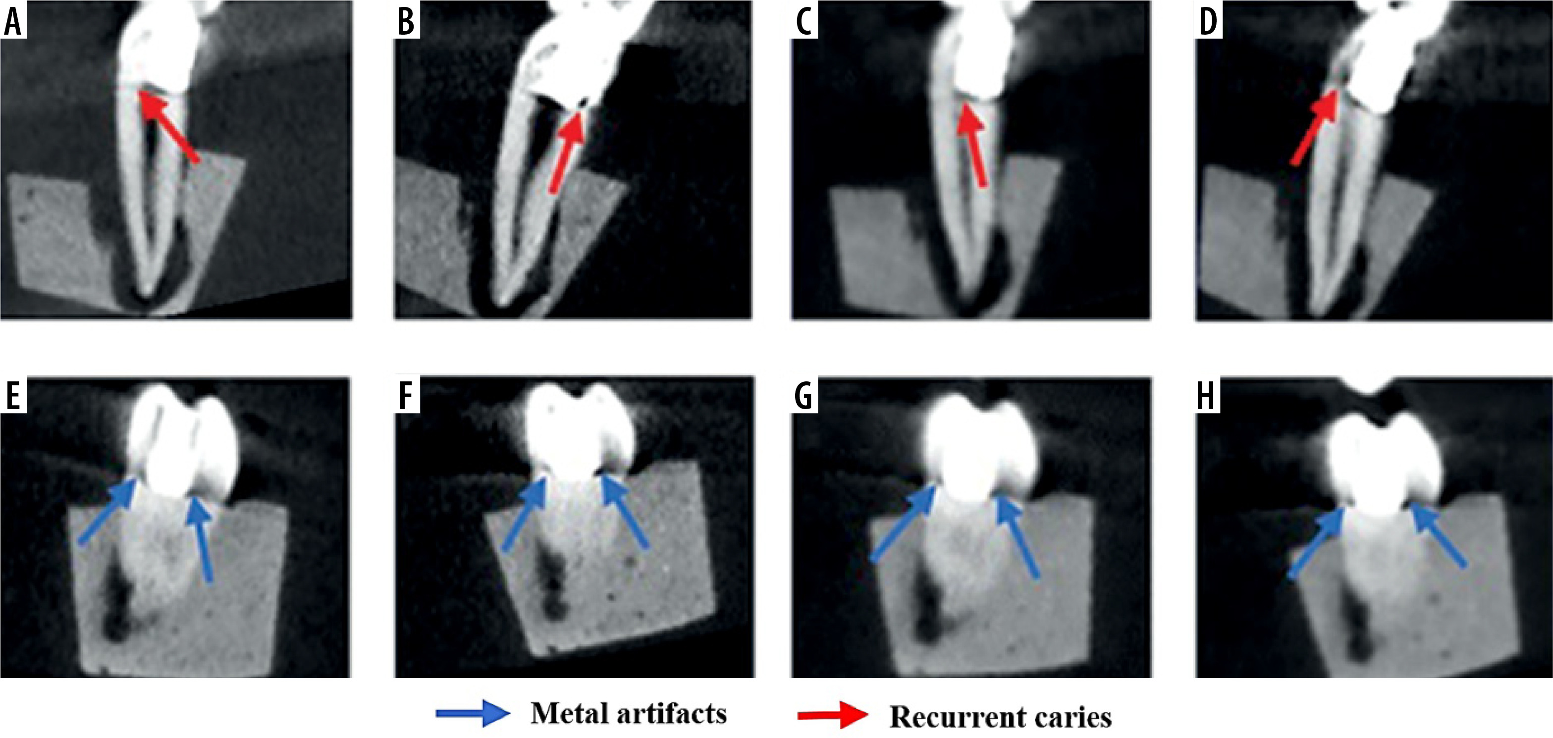
Figure 2
Cross-sectional cone-beam computed tomography images of composite Filtek Z250 restoration obtained by Giano NewTom. A) High-resolution (HIRes) setting without recurrent caries. B) Standard setting with metal artifact reduction (MAR) without recurrent caries. C) HIRes setting with MAR without recurrent caries. D) Standard setting without recurrent caries. E) HIRes setting with MAR with recurrent caries. F) HIRes setting with recurrent caries. G) Standard setting with MAR with recurrent caries. H) Standard setting with recurrent caries

Figure 3
Cross-sectional cone-beam computed tomography images of amalgam restoration obtained by Giano NewTom. A) HIRes setting with metal artifact reduction (MAR) with recurrent caries. B) High-resolution (HIRes) setting with recurrent caries. C) Standard setting with MAR with recurrent caries. D) Standard setting with recurrent caries. E) HIRes setting with MAR without recurrent caries. F) HIRes setting without recurrent caries. G) Standard setting with MAR without recurrent caries. H) Standard setting without recurrent caries
Images assessment
CBCT images were independently evaluated by 3 oral and maxillofacial radiologists with at least 10 years of experience in assessing caries on both CBCT and digital intraoral images; each observer evaluated all 76 images for the blocks. For evaluation purposes, the images were displayed on an EIZO S2000 FlexScan monitor with a resolution of 1600 × 1200 pixels (EIZO Nanao Corporation, Hakusan, Japan). Brightness, contrast, and zoom tools were used based on the visual needs of each evaluator. To better assess, the observers had access to the gamma parameters of each reconstructed tomographic image and the entire volume of scans. The blinded observers were previously trained on how to properly use the software in a special session. Observers created cross-sectional images. For each case, the observers were asked to state their diagnosis as follows: if they identified caries, they should report a positive sign (+); otherwise, a minus sign (–). The presence or absence of each simulated lesion was recorded in a form that could be used as the gold standard. Regarding the observation/measurement taken by the observers, a simplified 2-point scale method was used based on the study of Anbiaee et al. [14], due to the high confidence of the observers (95%) and many variables from the CBCT scan modes data to various restorative materials.
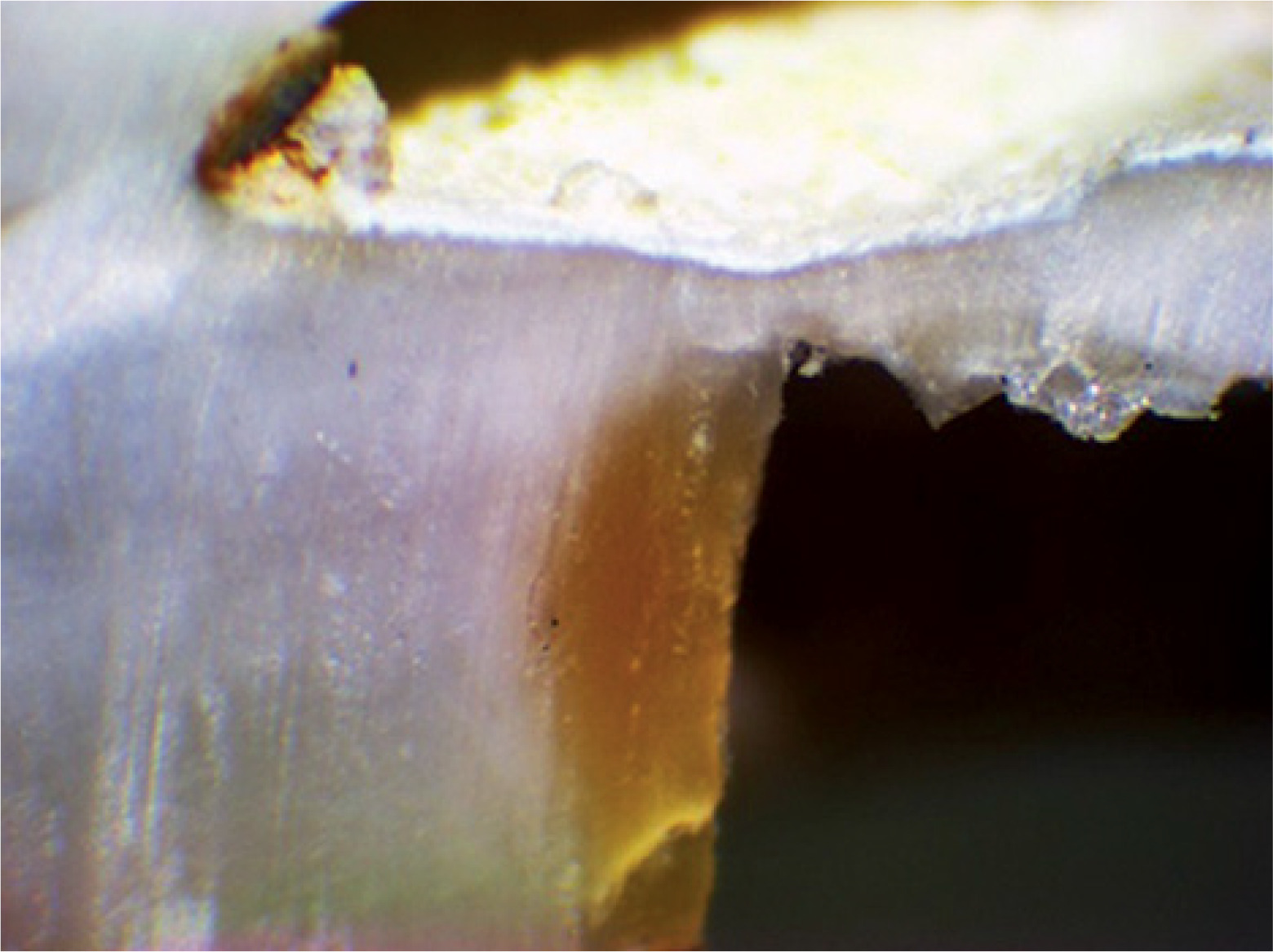
After preparing the radiographs, histological validation of the caries status was performed by sectioning each tooth mesiodistally at the restoration site using a diamond disk (Tizkavan, Tehran, Iran), and the sections were examined under a stereomicroscope (Dewinter, Milan, Italy) to ensure the presence of caries (Figure 4).
Statistical analysis
Diagnostic efficacy, including the area under the receiver operating characteristic curve (AUC), sensitivity, and specificity, were calculated to analyse the data for each setting. In this study, firstly, the diagnostic efficacy of 4 different configurations of CBCT scan modes (high- and standard-resolution, with and without the application of a MAR) and the significance of differences between pairs of these tests were compared. Then, AUC, sensitivity, and specificity were measured for different types of restorative materials under 4 different CBCT setting modes. The relationship between the Gray and AUC values was investigated using the Spearman correlation test. Cohen’s kappa was calculated to assess the reliability of the observers for each image set. Kappa values were interpreted on the basis of Landis and Koch’s guidelines [15]. The AUC and 95% confidence interval were calculated utilising SPSS software V19. The analyses were performed using MedCalc software (version 19.0.5). A significance level of p-value below 0.05 was considered for all analyses.
Results
Inter- and intra-observer agreement
Across all image modes, the validity for inter-observers and intra-observers was similarly obtained at 0.79. It should be noted that an observer’s agreement above 0.70 is substantial and confirms reliability [15].
Accuracy of CBCT scan modes with and without MAR
The AUC, sensitivity, and specificity values for each type of CBCT setting mode with and without the MAR option are presented in Table 1. According to the logistic regression model, AUC values were 0.5-0.7 for poor discrimination, 0.7-0.8 for acceptable discrimination, 0.8-0.9 for excellent discrimination, and more than 0.9 for outstanding discrimination [16]. The highest accuracy and sensitivity values were obtained for HIRes with and without MAR, whereas the lowest values were achieved for the standard mode. In addition, HIRes without the MAR option showed high specificity values. It should be noted that MAR does not affect the specificity values for both HIRes and standard modes; however, there were differences in the sensitivity and accuracy values between using and not using MAR for CBCT scan modes, particularly for HIRes mode.
Table 1
The sensitivity, specificity, and area under the receiver operating characteristic curve (AUC) values for 4 types of cone-beam computed tomography setting modes with and without the metal artifact reduction (MAR) option in the diagnosis of recurrent caries
| Setting type | Sensitivity (%) | Specificity (%) | AUC | p-value |
|---|---|---|---|---|
| HIRes MAR | 84.09 | 94.32 | 0.892 | < 0.001 |
| Standard MAR | 75.00 | 94.32 | 0.847 | < 0.001 |
| HIRes | 80.68 | 98.86 | 0.898 | < 0.001 |
| Standard | 7.27 | 93.18 | 0.852 | < 0.001 |
The results of the comparison of accuracy between HIRes and standard scan modes with and without the MAR option used to detect recurrent caries are represented in Table 2. The accuracy differences of the standard scan mode with the MAR option were significantly lower than those of HIRes with the MAR option (p = 0.018) and without the MAR option (p = 0.011), respectively. Also, without the MAR option, the accuracy of the standard mode was significantly lower than that of the HIRes (p = 0.020). In addition, the highest AUC values were found for the HIRes mode. Based on these results, the diagnostic efficacy of all imaging in the CBCT HIRes setting mode was higher than that in the standard mode.
Table 2
Comparison of accuracy between different types of cone-beam computed tomography setting mode with and without the metal artifact reduction (MAR) option
Accuracy of restorative materials in CBCT scan modes with and without MAR
Table 3 illustrates the AUC, sensitivity, and specificity for 5 types of restorative materials (Conventional composites [Filtek Z250, Gradia], flow composite [A2 Filtek Z350], glass ionomer [Fuji II LC], and amalgam [SDI-GS-80]) under different CBCT scan modes with and without the MAR. The specificity values were generally high for all filling materials under both CBCT scan modes with and without MAR option, whereas the sensitivity values were commonly low. When the specificity values increased (HIRes mode: composite, 100; Gradia, 95; flow, 93.75; glass ionomer, 100; amalgam, 82.35; Standard mode: composite, 100; Gradia, 90; flow, 93.75; glass ionomer, 100; amalgam, 88.24), the sensitivity values decreased (HIRes mode: composite, 100; Gradia, 94.74; flow, 93.75; glass ionomer, 94.74; amalgam, 31.25; Standard mode: composite, 94.44; Gradia, 94.74; flow, 81.25; glass ionomer, 84.21; amalgam, 12.50) using the MAR option for both CBCT modes. Moreover, without considering the MAR option, the specificity values for the HIRes scan mode (specificity: composite, 100; Gradia, 100; flow, 93.75; glass ionomer, 100; amalgam, 100;) were higher than those for the standard mode. Overall, amalgam was found to have the lowest sensitivity and AUC values among all modalities, resulting in a less accurate diagnosis of recurrent caries under amalgam restorations compared with other materials.
Table 3
The sensitivity, specificity, and area under the receiver operating characteristic curve (AUC) for 5 types of restorative materials under different cone-beam computed tomography scan modes with and without the metal artifact reduction (MAR)
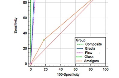
Figure 5 shows the receiver operating characteristic (ROC) curves based on the effect of the 5 restorative materials on the detection of recurrent caries under the 4 CBCT scan modes. As can be seen, a significant difference was found for all cases except for amalgam restorations under HIRes without the MAR option. Additionally, the accuracy of the standard CBCT mode with and without MAR in the amalgam group was significantly lower than that in the other 4 materials. There was also a significant difference between composite Gradia and glass ionomer (p = 0.025), the accuracy value of which was high for glass ionomer under HIRes scan mode with MAR.
Figure 5
The area under the curve based on 5 restorative materials for the (A) high-resolution (HIRes) metal artifact reduction (MAR) mode, (B) standard MAR mode, (C) HIRes mode, and (D) standard mode
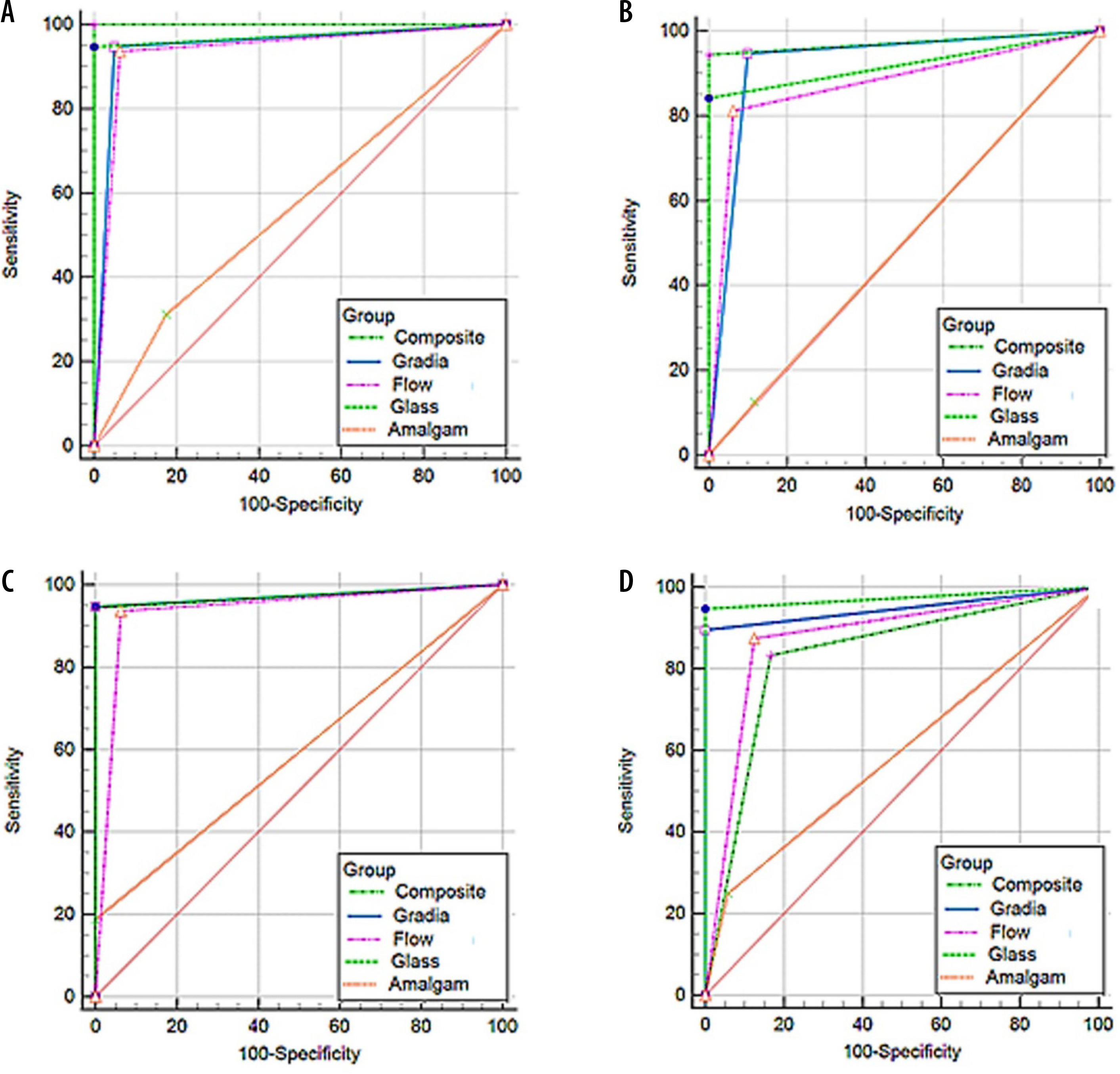
According to the above results, in each of the CBCT scan modes, no statistically significant differences were observed with and without the application of MAR for Gradia, flow, amalgam, and glass ionomer restorations, which resulted in a non-significant main effect on the accuracy and recurrent caries diagnosis, except in the case of composite restorations under standard CBCT scan mode.
Discussion
The present study aimed to compare the accuracy of different CBCT scan modes with and without the MAR option under 5 different filling materials in the detection of recurrent caries. Our results suggest a significant difference between the HIRes and standard-resolution scan modes. In this research, the highest AUC and sensitivity values were achieved for the HIRes mode with and without the MAR option, and the highest specificity was achieved for the HIRes mode without MAR. The results of AUC of 0.847-0.898 for the 4 types of CBCT scan modes showed an excellent ability to distinguish recurrent caries based on the logistic regression model [16]. The lowest AUC value (around 0.847) of the standard scan mode with MAR indicated that this CBCT mode may not be an ideal imaging technique for the detection of recurrent caries. However, in the survey by Kamburoglu et al. [17], no difference was found between different CBCT images with and without the MAR option to detect peri-implant and buccal periodontal. Besides, another study by Kamburoglu et al. showed that lower or higher AUC values of different methods are highly dependent on the depth of caries [1]. Moreover, the sum of sensitivity and specificity must be at least 1.5 for the tests to predict the presence or absence of disease [18]. In this study, all CBCT test results exceeded 1.5, which demonstrated that both high and standard resolution with and without MAR were valuable in detecting recurrent caries.
Although CBCT, as a promising technology, has shown the ability to detect caries in a satisfactory manner, its high radiation dosages remain a challenging step in various fields of dentistry [19,20]. Radiation exposure is a concern in radiographic caries diagnosis, and CBCT provides different doses based on the device, FOV, and other technical factors [12,21]. It should be noted that the use of CBCT for the diagnosis of recurrent caries is not a common approach, and it might not be the optimal imaging modality for caries assessment [22]. The present study investigated 4 types of CBCT scan modes with various exposure dosage levels to compare different setting types and determine the best CBCT scan mode for recurrent caries detection.
On the other hand, the presence of a high atomic number of restorative materials could create beam-hardening artifacts in CBCT images, which remarkably reduces the diagnostic image quality. During the evaluation of recurrent caries, artifacts usually appear as bright/dark lines adjacent to the margins of restorations, posts, and veneers. A metal object can absorb the X-ray beam, resulting in higher grey values near the object [23,24]. In other words, bright streaks caused by photon starvation could hide the carious lesion [25]. To solve these problems and reduce metal artifacts, various approaches have been developed.
One of the key benefits of CBCT is that it provides high spatial resolution images with relatively low radiation exposure. This is because CBCT uses a cone-shaped X-ray beam that rotates around the patient, capturing multiple images from different angles. These images are then reconstructed into a 3D volume using sophisticated algorithms. Although CBCT typically has lower spatial resolution than 2D images, its ability to capture volumetric data allows for better visualisation of complex anatomical features and relationships. This is particularly important in dental imaging, where even small changes in the structure of the teeth or surrounding tissues can have significant implications for diagnosis and treatment planning [25-27]. Although CBCT is generally considered safe, it involves exposure to ionising radiation. Radiologists and dentists should carefully weigh the risks and benefits of CBCT when deciding whether to use this imaging modality for a particular patient. In cases where the benefits outweigh the risks, CBCT can be an excellent tool for obtaining high-quality, detailed images of teeth and surrounding tissues [27-29].
MAR algorithms commonly use interpolation and smoothing methods to estimate and replace corrupted data around metal objects. However, the smoothing process in these algorithms can sometimes result in the loss of fine details in the image. This means that structures near metal objects may appear less sharp or distinct. Balancing artifact reduction with image sensitivity can be a tricky task for radiologists. They may need to fine-tune MAR parameters such as interpolation levels and smoothing to preserve critical details while reducing artifacts. Radiologists should consider the trade-off between reducing metal artifacts and maintaining image sharpness when selecting MAR algorithms. In some cases, it may be necessary to selectively apply MAR to specific regions of interest rather than the entire image [30-32].
Limited studies have investigated the effect of MAR on recurrent caries detection under different types of restorations. Because the detection of recurrent caries by CBCT under amalgam restorations is known to be a complicated process in the presence of artifacts, the present research investigated 5 types of restorative materials under 4 CBCT scan modes with and without artifact reduction. Images acquired of different types of CBCT scan modes under various restorative materials presented the lowest specificity values for HIRes and standard scan modes with the MAR option in the amalgam group, as well as the lowest sensitivity and accuracy values for HIRes and standard scan modes without the MAR option, in which a more negative effect of artifacts was expected. This result may be related to beam hardening, which creates dark zones adjacent to the restorative materials. Artifacts mostly occur in the presence of metallic restorations, leading to reduced accuracy, sensitivity, and specificity of CBCT [33,34]. In this study, there were differences in specificity and sensitivity values between HIRes and standard CBCT scan modes. In the amalgam group, the specificity and sensitivity were too low for both HIRes and standard modes with the MAR option. However, the specificity tends to increase in the standard scan mode, which may be due to the reduction in the number of surfaces incorrectly detected as decay and beam-hardening artifacts by the MAR algorithm. Moreover, it has been found that the number of beam-hardening artifacts in CBCT scans can vary between different devices [35,36].
The composite restoration groups used in this study showed identical levels of specificity in both HIRes and standard mode with MAR, whereas the standard scan mode had the highest sensitivity values. Similarly, Cebe et al. [12] achieved the same result in composite groups. Since radio-opacity is a key feature of restorative materials, the difference in values obtained might be related to the level of radiopaque content, which is highly dependent on the manufacturers to improve the radiopacity degree of products [12]. Overall, in the case of the standard scan mode, the accuracy value in the amalgam group was significantly lower than that of the Z250 composite, and there was no significant difference between other materials. Kulczyk et al. [37] studied the effect of amalgam fillings on the detection of proximal caries by CBCT using a NewTom 3G scanner (0.25 mm voxel size, 9 × 9 cm FOV). They reported that lower sensitivity, specificity, and accuracy values were achieved for detecting carious lesions in enamel. In the study carried out by Mattson et al. [38], it was found that the detection rate of recurrent caries in intraoral films was high in the vicinity of the radiopaque composite and low when adjacent to the radiolucent composite.
ROC analysis is mostly used to evaluate the diagnostic performance of different imaging systems [39]. The comparison results of the 4 CBCT scan modes showed that the HIRes scan mode performed better than the standard scan mode, with the highest AUC, specificity, and sensitivity values. It has been suggested that the higher-resolution scan mode of CBCT images could increase the accuracy of caries diagnosis [40]. Additionally, the AUC for the HIRes CBCT scan mode with the MAR option demonstrated excellent diagnostic performance, especially in the composite group. Furthermore, we found that the caries detection accuracy in amalgam was significantly lower than that of Z250 composite and glass ionomer in both CBCT scan modes, regardless of whether artifact reduction was used. Overall, these findings suggest that evaluating CBCT scan modes under different filling materials generally increases the possibility of correct diagnosis of recurrent caries using the MAR option.
The results of a study by Sausa et al. [41] showed that CBCT images in higher resolution under composite restorations performed better than conventional digital radiography in detecting recurrent caries, which was similar to the results of the present study. Compared with our amalgam group, they obtained a lower sensitivity (0.27 for HIRes). Although intraoral radiography showed high spatial resolution in various cases, CBCT as a 3D technique can prevent super-imposition and reveal more caries. In the study conducted by Charuakkra et al. [42], 2 CBCT systems (Pax-500ECT and Promax 3D) were used, and the AUC values for Pax-500ECT and Promax 3D were 0.995 and 0.978, respectively.
The current study observed substantial intra- and inter-examiner validity for all groups of restorative materials in both CBCT scan modes. It should be noted that if the observers suspected a Mach band effect error, they ruled it out. However, many factors can influence the level of agreement, including the observer’s experience, evaluation conditions, access to software tools, and the method of reading multiplanar images. Distinguishing caries from artifacts can sometimes be challenging for observers, leading to difficulties in interpreting CBCT images to detect recurrent caries [12].
Overall, the results of this study suggest that using the HIRes CBCT scan mode with the artifact reduction option can increase the accuracy and sensitivity of detecting recurrent caries, especially in composite groups. Therefore, when a diagnosis of caries is suspected in clinical practice, the use of the HIRes scan mode should be considered to improve the detection of recurrent caries under different restorative materials, regardless of MAR application. Contrary to our findings, Cebe et al. [12] found that using a MAR option in conjunction with CBCT scans can improve the accuracy of approximal caries detection. Additionally, our study revealed that the specificity values were generally high for all filling materials under both CBCT scan modes with and without the MAR option, whereas the sensitivity values were commonly low. This finding indicates that CBCT imaging may not be the most suitable tool for detecting early-stage recurrent caries. Therefore, clinical practice should also consider other diagnostic methods, such as visual inspection and radiographic examination, to complement CBCT imaging.
The main limitation of this in vitro study is that because of laboratory conditions, we mounted extracted teeth, and soft tissue reconstruction may lead to some errors and artifacts that complicate caries detection [24]. It is suggested that different and more common restorative materials be evaluated in each geographical region with other CBCT systems in future studies. Finally, it should be noted that MAR algorithms may degrade image quality and hamper the visualisation of crucial radiological signs [43,44].
Conclusions
CBCT potentially has a high diagnostic performance in recurrent caries detection. In particular, the use of the HIRes CBCT scan mode resulted in increased accuracy and sensitivity for recurrent caries detection compared with the standard CBCT scan mode, irrespective of MAR application. On the other hand, the accuracy in detecting recurrent caries was lower in the amalgam group compared with other restorative material groups. It should be noted that the use of the HIRes scan mode should be considered when caries diagnosis is suspected.