Introduction
The coronary sinus is the final region of the cardiac venous system. It lies on the backside of the heart, between the left atrium and left ventricle. The coronary sinus starts at the junction of the great cardiac vein, and sometimes the oblique vein of the left atrium (vein of Marshall); however, this location is often difficult to define. Its ostium finds to the right atrium [1,2]. The role of the coronary sinus is to collect venous blood and discharge it to the heart chambers [3,4]. Its opening is often covered by an incomplete membrane called the Thebesian valve [5,6]. Several types of coronary sinus have been described in the literature [3,7]. The coronary sinus appears to act as a buffer in several pathologies, such as heart failure or coronary artery disease; hence, the volume of this structure may be important [8]. So far, we only know measurements of the volume of the coronary sinus from autopsy studies [9]. Modern imaging techniques such as computed tomography can help in the assessment of this parameter in a vitro manner, but there is no comprehensive research on this topic so far. Hence, we decided to develop a methodology for measuring the volume of the coronary sinus in multi-detector computed tomography (CT) and to try to apply it in practice.
Material and methods
CT scanning
CT images of 49 patients (22 men) the quality of which were accepted for clinical evaluation were included in this trial. Scans of 3 patients were excluded due to clinically unaccepted image quality including artifacts. The average age of the patients was 70.08 ± 13.6 years (range 31-86 years). The main reason for performing CT in all patients was coronary artery disease suspicion. Scanning with retrospective ECG-gating was performed during a breath-hold using a Toshiba Aquilion 64 with a collimated slice thickness of 0.5 mm, according to the standard protocol for coronary arteries [10]. The best mode option was used in all cases; the helical pitch was 12.8, and the rotation time was 0.4 s. The tube voltage was dependent on the patient’s weight. An average of about 80 ± 20 ml of non-ionic contrast was administered to each patient in the study. In some cases (heart rate > 65 bpm), metoprolol succinate was administrated intravenously, unless contraindicated.
Post-processing
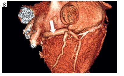
The volume of coronary sinus and other data measurements were performed using Vitrea 2 (Vital Images, Minnetonka, MN, USA) workstations. Multi-planar reformatted reconstructions and three-dimensional (3D) volume renderings were used. Characterisations of coronary sinus measurements included density in Hounsfield units (HU) and volume in cubic centimetres. To standardise the measurements, they were all performed to the place where the vein of Marshall reached the coronary sinus [11]. In cases of loss of the vein of Marshall, the first lateral vein was used as the border between the coronary sinus and the great cardiac vein. The organ volume measurement function of the Vitrea 2 workstation was used to automatically measure and display the volume of objects in CT scans (Figure 1A). In some cases, manual correction was performed. The analyses were performed by 2 experienced researchers. Quality of visualisation of coronary sinus was also performed based on a previously invented and published method [11].
Statistical analysis
Statistical analysis of the obtained data was performed using MedCalc software, version 22.021 (MedCalc Software Ltd, Ostend, Belgium). Continuous data are presented as the mean ± standard deviation. Correlations were made using the Pearson method. The results were considered to be statistically significant at p < 0.05.
Results
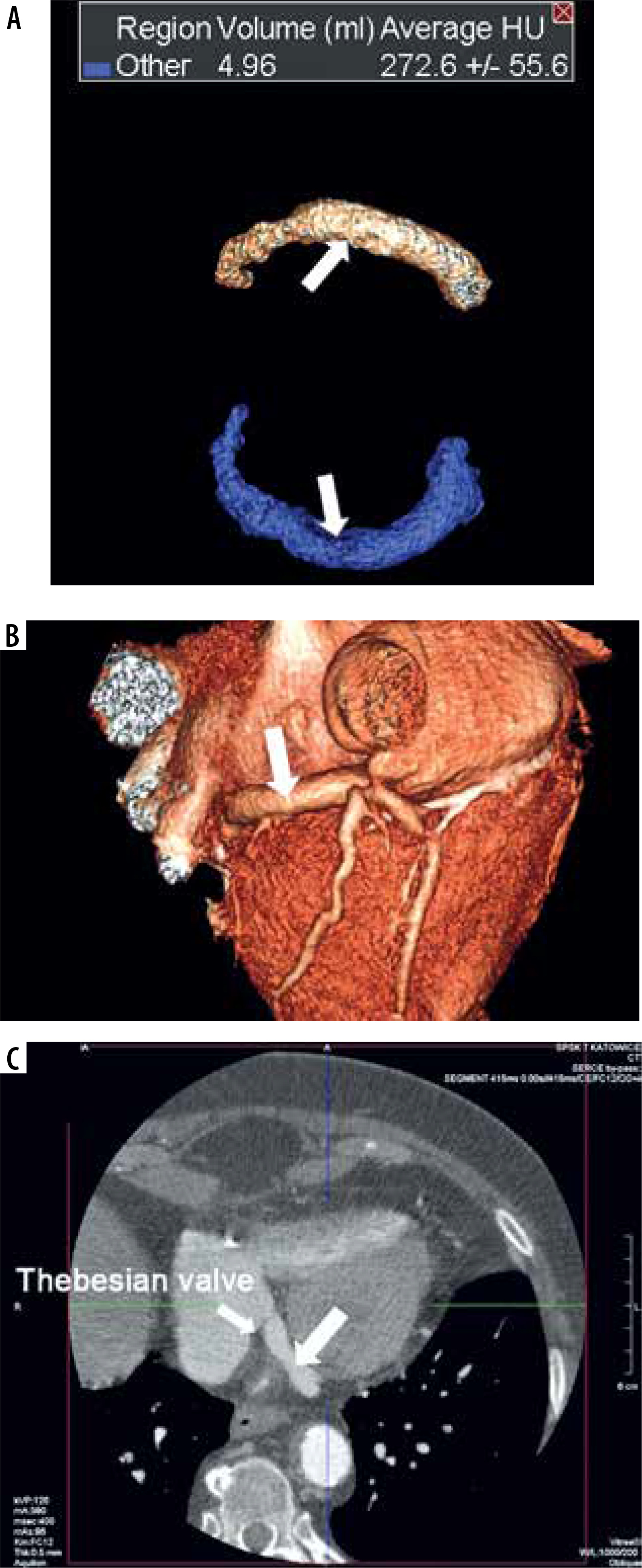
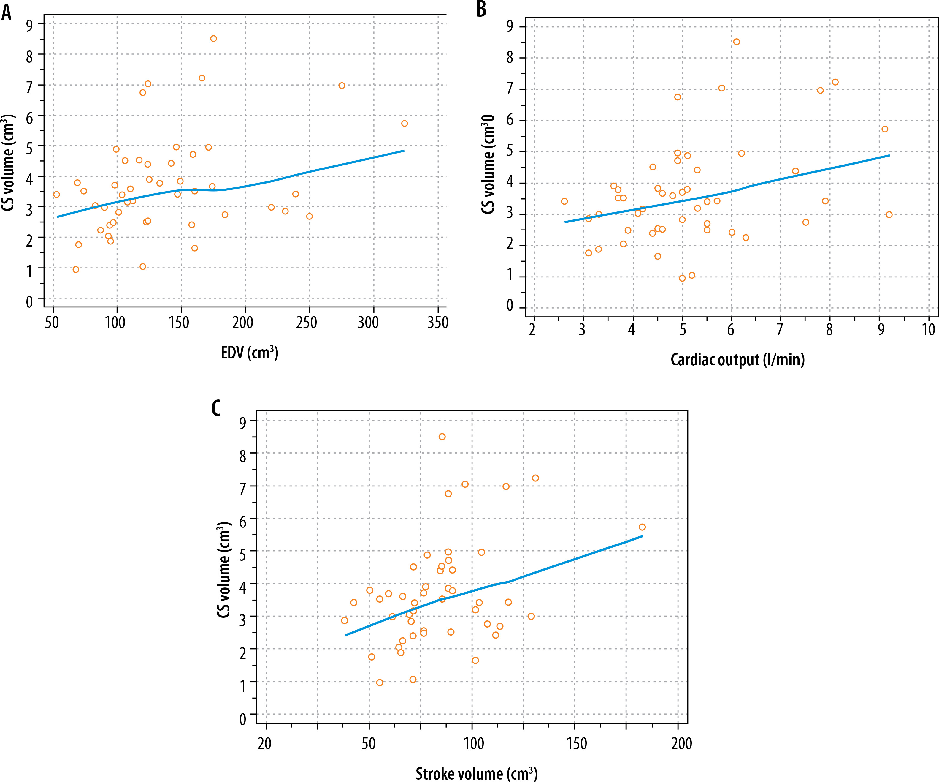
Haemodynamic characteristics of the included patients are presented in Table 1. Coronary sinus volumes measured in the research varied from 0.96 to 8.52 cm3. The average volume was 3.71 ± 1.64 cm3. An example of visualisation and measurement is presented in Figure 1. There are a few significant correlations between coronary sinus volume and selected haemodynamic measurements: end diastolic volume (r = 0.33; p = 0.02), stroke volume (r = 0.39; p = 0.01), and cardiac output (r = 0.40; p = 0.01). These correlations are graphically presented in Figure 2. Typical 2D measurements were as follows: angle of entrance to the right atrium was 104.5 ± 11.23°, length of coronary sinus ostium was 10.37 ± 2.5 mm, and the average diameter one cm from the CS ostium was 10.38 ± 2.3 mm. Because measurement of the 3D shape of the coronary sinus relates to its density, the average value was 263.5 ± 90.29 HU (minimal value was 26.3 HU, maximum value 447.9 HU). In most cases the quality of visualisation was good – the average was calculated as 4.16 ± 0.87 cm3 (range 2-5 cm3). The Thebesian valve was present in 22 cases (44.9%); however, no statistical relationship between the presence of the Thebesian valve and coronary sinus was observed.
Table 1
Haemodynamic characteristics of the included patients
Discussion
There are several options for imaging the coronary sinus. Some of them are post-mortem tests. In vivo imaging of the coronary venous system has been available for several years [11]. Among those methods, multidetector cardiac CT offers the highest accuracy, preferably with retrospective gating [12-15]. The coronary sinus in CT has been described many times. In light of reports that the coronary sinus and the great cardiac vein play a role in various cardiac pathologies, we hypothesised that the volume of the coronary sinus may be important. In our earlier report we documented that the CABG (coronary artery bypass graft) procedures can change the distribution of pressures within the arterial and venous vessels, and we believe that coronary artery diseases may be target pathologies for the evaluation of coronary sinus volume [16]. Similarly, the statistically higher number of veins in patients with heart failure may suggest an association between a failing heart and cardiac venous retention [8].
There is only one study that comprehensively measured the volume of the coronary sinus along with other measurable parameters: Silver and Rowley studied 50 human coronary sinuses with normal and increased cardiac mass to determine various functional anatomical features of the coronary sinus and differences between normal and increased volume hearts [9]. The mean coronary sinus volume was 1.26 ± 0.45 cm3, while in our research it was larger, at 3.71 ± 1.64 cm3. We believe the difference is caused by different evaluation methods. In our research we use in vivo diagnostic methods – CT contract enhancement, while and Silver and Rowley used post-mortem cadaveric hearts. Another potential reason for the discrepancy is the determination of the strict border of the coronary sinus/great cardiac vein. In the cited paper, in hearts of weight from 365 to 675 g, the Thebesian valve covered the ostium an average of 26% and the average volume of the coronary sinus was increased to 1.76 ± 0.73 cm3 (p < 0.005). In our study, we did not observe any statistical correlation between coronary sinus volume and myocardial mass (r = 0.08; p = 0.59).
The biggest problem for us during the design of this study was to determine the borders of the coronary sinus as precisely as possible. If the oblique vein of the left atrium, called the vein of Marshall, was available, we considered this place as the beginning [17,18]. In the case of its absence, which is quite common, we assumed that the first lateral vein of the heart from the mouth of the coronary sinus constitutes the boundary. The latter solution constitutes a certain type of contract and seems to be the only one that can be applied.