Introduction
Nephrolithiasis, or renal calculi, can be considered as the consequence of crystallization and aggregation of highly concentrated urinary components, and they are caused by a disruption in the balance between solubility and precipitation of salts in the urinary tract and in the kidneys. There are basically 2 main categories of stones, calcium stones and non-calcium stones. The most common chemical compositions are calcium oxalate (70%), calcium phosphate (20%), uric acid (8%), and cystine (2%).
The lifetime prevalence of urinary tract stones has been estimated at 10-14% [1]. The morbidity associated with urolithiasis includes colic pain and obstructive uropathy, which can lead to renal failure and severe urinary tract infections, leading in turn to pyonephrosis and septic shock.
Knowledge of the composition of urinary tract stones is fundamental in planning appropriate treatment and preoperative patient evaluation, and this information influences both treatment plans and recurrence prevention. For example, stones composed of cystine or calcium oxalate monohydrate have a firm composition that may limit the success of extracorporeal shock wave lithotripsy. These stones may be more effectively treated with percutaneous nephrolithotomy (PCNL) [2].
Systemic and familial metabolic nephrolithiasis may be suspected in patients with certain stone composition, and specific dietary and medical measures to reduce the risk of recurrence can be offered if the type is known [3].
Until recently, the diagnosis of renal stone disease was mainly done through urine analysis and formal stone analysis once the stone is removed. However, dual-energy computed tomography (DECT) not only has the ability to detect stones with sensitivity close to 100% but also offers a novel technique of analysing the composition of the stone during routine CT scan [4]. The majority of renal stones are calcium oxalate stones and account for up to 75-80% of renal stones. Struvite (magnesium ammonium phosphate), calcium phosphate, uric acid, cystine, and mixed stones make up the remaining 20-25% [5]. Although renal stones can be picked up in X-ray and USG with varying sensitivity, they play no role in the actual assessment of the composition of the calculi.
With approximately 50% of patients expected to present with recurrence of renal calculi over 10 years from the initial presentation, it is of paramount importance to make every attempt to ensure that recurrence is negligible. It is in this area that knowledge of the composition of the calculi plays a significant role [6]. Renal calculi analysis is essential part of the workup in patients with established renal calculi disease with a moderate and high risk of recurrence.
For several decades 24-hour urine collection has been traditionally used to determine the composition of calculi in the urinary tract. However, this test can be inaccurate and is time consuming [7]. Thus, stone fragment analysis is an important approach in the management of renal calculi disease; however, it depends on the actual availability of calculi, which at times is difficult when small stones have been expelled. During several urological procedures the stones are crushed and are washed away into flushing solutions. The 3 most common ex vivo techniques for stone analysis are X-ray diffraction, infrared spectroscopy, and polarization microscopy. These methods of chemical analysis of the urinary calculi are costly and time consuming.
Dual-energy CT, by facilitating low- and high-energy scanning during a single acquisition, has the inherent capability to differentiate materials that have similar electron densities but varying photon absorption. Our aim in this study was to preoperatively assess the composition of urinary tract stones with dual-energy CT and its comparison by using postoperative in vitro infrared spectroscopy analysis as the reference standard.
Material and methods
This was a prospective study done for a period of 9 months in all patients who underwent dual-energy CT examination for the evaluation of urinary tract stones. The patients included in this study underwent percutaneous or ureteroscopic stone extraction and infrared spectroscopy validation of the collected stones in our hospital. Informed consent was obtained from all the participants.
All examinations were performed with a third-gene-ration, dual-source, dual-energy CT scanner (Siemens Somatom FORCE scanner). The scan was performed using 100 kVp and 150 kVp. The scan was covered from the upper pole of the kidney to the pubic symphysis. The images were reconstructed with a slice thickness of 1 mm and were sent to a syngo.via workstation. The images were analysed in the syngo.via workstation, and the components of the stones were identified. The software displayed calcium stones in blue and non-calcium (uric acid) stones in red.
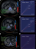
The urinary stone CT density was measured using a region of interest equal to 50 % of the maximal diameter of each stone. With Dual energy CT, renal stone attenuation at low and high kVp was attained and the attenuation ratios was measured. We characterized the stones based on the attenuation ratio as follows: Less than 1.1 for uric acid, 1.1-1.24 for cystine, and greater than 1.24 for calcified stones (Figure 1) [8]. Struvite stones had attenuation ratios that overlapped with calcified stone ratios and thus could not be assessed reliably [9]. For each patient, the location (kidney, ureter, or bladder) and the maximal dia-meter of stones were also evaluated. These patients underwent percutaneous or ureteroscopic non-destructive stone extraction and were sent for analysis of the composition using Fourier infrared spectroscopy.
Figure 1
Dual-energy images and graph demonstrating different composition of renal stones. Uric acid calculus (A-B), calcium stone (C-D), and cysteine stone (E-F)

Statistical methods
Descriptive analysis was carried out by mean and standard deviation for quantitative variables, and frequency and proportion for categorical variables. The association between stone type as assessed by DECT and stone type as assessed by laser IR was estimated by cross tabulation and comparison of percentages. Chi-square test/Fisher’s exact test was used for statistical analysis. P-value < 0.05 was considered statistically significant. IBM SPSS version 22 was used for statistical analysis.
Results
Our study showed that the prevalence of renal stones was more common in the male population (38 out of 50, 76%) than in the female population (12 out of 50, 24%). The mean age for the occurrence of renal stone was found to be 47 years (between 43 and 50 years). Of 50 patients, 38 had stones in the kidney, 5 in the ureter, and 3 in the bladder. The remaining 4 patients had stones in the kidney and ureter. In our study, the stone diameters varied between 4 and 40 mm (mean diameter 14 mm).
The mean attenuation DE ratio value of the renal stones in this study population was 1.57 ± 0.25. Out of 50 patients, in 39 the stone type was predicted as calcium-containing stones on DECT, which were confirmed as calcium stones on laser spectroscopy (90%). In 7 patients the stone type was predicted as containing uric acid on DECT and was confirmed as uric acid stone on laser spectroscopy (100%). In 4 patients, the stone type was predicted as cystine stone and turned out to be calcium stones on laser spectroscopy (Table 1). On in vitro infrared spectroscopy analysis, the most common stone type in the sampled patients was calcium oxalate (86%). Thus, the positive predictive value for DECT to accurately assess the chemical composition of the sampled renal calculi, as confirmed by infrared spectroscopy analysis, was found to be 92% (Table 2). A comparison of the DECT and IR spectroscopy results is shown in Table 3.
Table 1
Stone types as predicted by computed tomography (CT) and infrared spectroscopy (IS) in the study population (N = 50)
| Stone type | Modality | |
|---|---|---|
| CT, n (%) | IS, n (%) | |
| Calcium | 39 (78) | 43 (86) |
| Cysteine | 4 (8) | 0 |
| Uric acid | 7 (14) | 7 (14) |
Table 2
Descriptive analysis of correlation of dual-energy computed tomography findings with infrared spectroscopy (IS) findings in the study population (N = 50)
| Correlation with IS findings | n (%) |
|---|---|
| Yes | 46 (92) |
| No | 4 (8) |
Discussion
The composition of the imaged volume of tissue is determined by the differing attenuations of X-ray beams, which are received by the detectors and then computer processed and reconstructed to produce virtual slices of that particular volume of tissue. Within each slice we can confidently distinguish between bone, air, fat, soft tissue, fluid, etc. However, tissues that are of similar density and atomic number look identical on conventional CT; for example, calcium oxalate and uric acid renal stones are indistinguishable. This is where the use of dual-energy CT takes a step further into the realm of tissue characterization. With these techniques 2 X-ray tubes of different tube voltage potentials, usually 100 kVp and 150 kVp, image the same tissues, in our case a renal stone.
Two stones of differing atomic composition will demonstrate differences in attenuation in 2 energy levels, and this can be analysed using post-processing techniques to provide information about tissue composition beyond that obtainable with a single-energy technique. Initial studies, both in vitro and in vivo, have been promising, with dual-energy CT correctly identifying renal calculi composition with 92-100% accuracy [10]. Several studies have showed the ability of dual-energy CT to accurately differentiate between the major pure-stone groups in vitro. Limited in vivo studies have proven the accuracy in differentiation between uric acid and non-uric acid groups [8,11-13].
Dual-energy CT as a method of non-invasive renal stone characterization is an exciting and novel development with far-reaching implications in the investigation and diagnosis of patients and subsequently the management of renal stone disease while obviating the need for time-consuming or invasive procedures. The characterization of various stones is based on the dual-energy ratio and X-ray attenuation or HU.
Nephrolithiasis in the current study was more common in males (76%) than in females. The age of our selected patients ranged from 35 to 50 years with a mean of 47 years.
Uric acid stones contain low-molecular-weight elements (carbon, nitrogen, oxygen, and hydrogen), which show increased attenuation in high-kVp images. Non-uric acid stones (calcium oxalate, hydroxyapatite, and struvite stones) have high-molecular-weight elements like calcium, phosphorus, and sulphur, which show decreased attenuation in high-kVp images [14]. This results in a decreased dual-energy attenuation ratio for uric acid stones and an increased attenuation ratio for non-uric acid stones.
In our study, we followed the dual-energy attenuation ratio as follows: less than 1.1 for uric acid stones; ratios of 1.1-1.24 for cystine stones; and ratios of 1.25-2.4 for calcified stones [15]. In our study, DECT accurately differentiated uric acid stones (7 stones, representing 14%) from non-uric acid calculi (43 stones, representing 86%), which agreed with previous studies in the literature [10-14,16].
We found in our study that the most common type of renal stones in DECT was calcium oxalate, seen in 39 patients (78%), followed by uric acid stones in about 14% and cystine stones about 8%. The results of our study agreed with those of the study by Ferrandino et al. [17]. In their study they also found calcium oxalate to be the most common renal stone (56%) and uric acid stones as the second common (28%) followed by cystine stone (16%).
We compared the composition predicted by DECT with the results of laser spectroscopy ex vivo stone analysis after PCNL stone extraction. In our study, in vivo characterization of kidney stones was achieved using dual-energy CT correctly in 92% of patients (46 out of 50 patients). In the remaining 4 patients, DECT characterized the stones as cystine stones, but they were shown to be calcium stones on IR spectroscopy. The results were similar to those of the study done by Manglaviti et al. [14]. In a study done by Spek et al. the DECT results and postoperative infra-red spectroscopic analysis had an agreement of 98.4% [18]. In another study done by Hidas et al. the diagnostic accuracy of single-source, dual-energy CT in characterizing renal stones was 82% [15].
The clinical management of urinary tract stones depends on the location, size, and number of calculi as well as their chemical composition. Urinary calculi that consist primarily of uric acid can be treated with oral medications (urine alkalization). Cystine, calcium oxalate, and brushite-based stones are less fragile than other types of calculi and are unlikely to benefit from extracorporeal shock wave lithotripsy (ESWL), which can be expensive if repeated treatments are necessary, and in some cases it results in renal haemorrhage and fibrosis [19]. These types of stones are better treated with ureteroscopy of percutaneous nephrolithotomy. In our study, we were able to differentiate uric acid from non-uric acid stones in all patients.
There were some limitations in our study. First, our patient population was relatively small; this was due to the inclusion requirement that all study patients subsequently undergo PCNL. Another limitation was that stones smaller than 3 mm were not included in our study. Previous work has shown that accuracy begins to decline in such small stones because of partial volume averaging. In our study we did not include struvite stones because of their mixed composition (apatite, oxalate, and calcium phosphate).
Conclusions
The third-generation DECT scan had 100% accuracy in differentiating uric acid stones from non-uric acid stones in our study. Because the treatment varies depending on the chemical composition of stones, identification of specific chemical componentsw is very important, and this can be accurately done by DECT.


